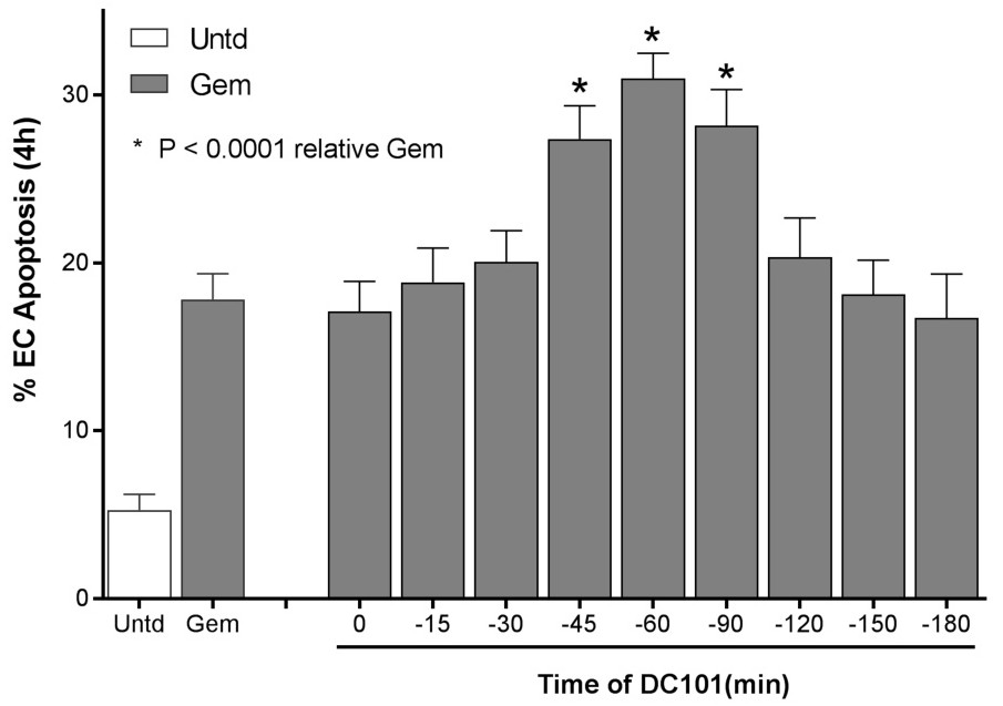
Fig. 2. A 45 min pre-treatment window defines sphingolipid-based anti-angiogenic chemosensitization. 1x10⁶ MCA/129 fibrosarcoma cells were implanted into the right flank of sv129/Bl6JAX mice. When tumors reached an average of 100mm3, DC101 (1600 μg/mouse i.v.) was delivered at the indicated times preceding Gemcitabine (Gem; 240 mg/kg i.p.). Mice were sacrificed at 4h after Gem, and 5-μm thick tumor sections were double stained with TUNEL to detect apoptosis and MECA-32 Ab to identify endothelial cells. Data (mean±95% Cl) derive from ~2000 endothelial cells (ECs)/group collated from 3 mice each.